Microplate formats underpin nearly every high-throughput screening (HTS) workflow, influencing assay design, data quality, and overall screening throughput. As laboratories scale biochemical, cell‑based, and reporter assays, selecting between 96‑, 384‑, and 1536‑well microplate formats becomes a critical decision.
These microplate formats differ not only in well density but also in liquid handling requirements, signal‑to‑noise performance, and compatibility with automation. Understanding these distinctions enables more efficient assay development and optimization of screening throughput.
This article provides a comparison of microplate formats, outlining the characteristics, advantages, and limitations of 96‑, 384‑, and 1536‑well plates for cell-based and biochemical HTS assays.
Microplate format fundamentals and HTS plate density
Microplate formats are defined by well count, working well volume, and working well surface area (Table 1).
Table 1: The typical characteristics of common microplate formats
|
Format |
Well count |
Typical well working volume |
Typical working surface area |
Use cases |
|
96-well |
96 |
25-340 μL |
34mm2 |
ELISAs, cell‑based assays, kinetic biochemical assays |
|
384-well |
384 |
15-145 μL |
10mm2 |
HTS biochemical assays, reporter assays |
|
1536-well |
1536 |
3-10 μL |
2mm2 |
Ultra‑HTS, miniaturized biochemical screens |
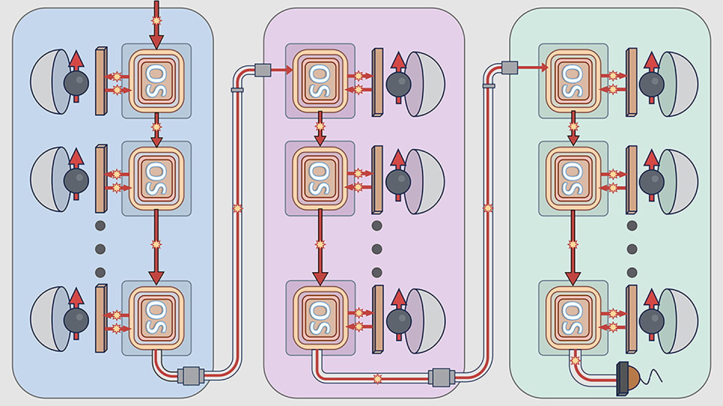
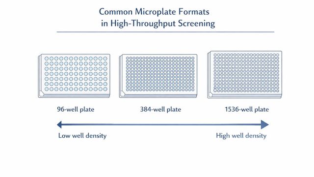
Microplate wells can come in round, square, or rounded-square geometries. These parameters directly influence assay miniaturization, reagent consumption, and throughput. HTS plate density has increased steadily over the past two decades, driven by improvements in liquid handling precision and detection sensitivity (Figure 1).

Figure 1: The common microplate formats in high-throughput screening. Credit: AI-generated image through Microsoft Copilot (2026).
Key well density considerations
- Increasing well density reduces working volume, which can amplify evaporation and edge effects.
- Higher density formats require improved pipetting precision, especially below 10 µL.
- Detection sensitivity must be sufficient to maintain the assay window at reduced volumes.
Standards such as the ANSI/SLAS microplate specifications provide dimensional guidelines to ensure compatibility with automation systems.
Assay performance across microplate formats
Assay class strongly influences the suitability of each microplate format. Biochemical assays often tolerate miniaturization well, whereas cell‑based assays may require larger volumes to maintain viability and reduce variability.
Biochemical assays and miniaturization efficiency
Biochemical assays—such as enzyme kinetics, binding assays, and reporter‑based biochemical readouts—are typically the most amenable to high‑density formats.
Why biochemical assays scale effectively:
- Homogeneous reaction conditions reduce variability at low volumes.
- High signal‑to‑background ratios can be maintained with optimized reagent concentrations.
- Miniaturization significantly reduces reagent cost per data point.
Studies have shown that many biochemical assays maintain Z’‑factor performance above 0.5 even when miniaturized to 1536‑well volumes.
Considerations for 384‑ and 1536‑well formats:
- Dispensing accuracy below 1 µL requires acoustic or non‑contact liquid handling.
- Mixing efficiency becomes a limiting factor in ultra‑low volumes.
- Edge wells may show increased evaporation unless humidity control is implemented.
Cell‑based assays and format constraints
Cell‑based assays introduce additional constraints due to cell viability, adherence, and sensitivity to microenvironmental changes.
96‑well plates remain widely used for cell‑based assays because they offer larger volumes that buffer against evaporation, adequate surface area for adherent cell growth, and higher tolerance for manual handling.
384‑well plates are increasingly used for high‑content imaging and reporter assays, provided that cell seeding uniformity is tightly controlled, liquid handling systems can dispense low volumes without disturbing cells, and incubation conditions minimize edge effects.
1536‑well plates are less common for cell‑based assays due to their limited surface area for cell attachment, increased sensitivity to evaporation and temperature gradients, and challenges in maintaining uniform cell distribution.
However, some ultra‑miniaturized cell‑free reporter assays and luminescent readouts perform well in 1536‑well formats.
Screening throughput optimization in microplate formats
Throughput is influenced by plate density, assay cycle time, detection speed, and automation compatibility. Selecting the appropriate microplate format requires balancing throughput with data quality.
A single 1536-well plate contains 4 times as many wells as a 384-well plate, or 16 times as many wells as a 96-well plate. This dramatically increases screening capacity. For example, screening 100,000 compounds requires ~1,042 plates in 96‑well format. The same library requires only ~65 plates in 1536‑well format.
This reduction in plate count decreases incubator space requirements and accelerates screening timelines.
Liquid‑handling precision and automation compatibility
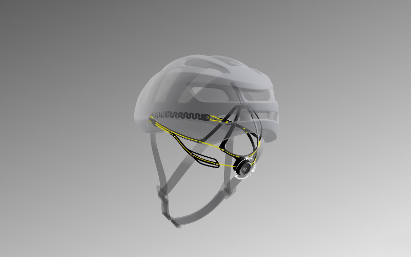
Automation plays a central role in enabling high‑density microplate formats, but factors including pipetting precision and detection sensitivity must be considered (Figure 2).

Figure 2: Automation considerations for common microplate formats in HTS. Credit: AI-generated image through Microsoft Copilot (2026).
Key automation considerations:
- Pipetting precision: Sub‑microliter dispensing requires acoustic droplet ejection or high‑precision non‑contact systems.
- Plate handling: ANSI/SLAS‑compliant plates ensure compatibility with robotic arms and stackers.
- Evaporation control: Humidity chambers and plate seals reduce variability in 384‑ and 1536‑well formats.
- Detection sensitivity: Miniaturized assays require sensitive luminescent or fluorescence detection systems.
While 96‑well plates can be used manually, 384‑ and 1536‑well formats are typically automation‑dependent for reproducible performance.
Assay window and data quality considerations
Assay robustness is often quantified using the Z’‑factor. Miniaturization can reduce assay window (the ratio between signal and background) due to:
- Increased surface‑to‑volume ratio
- Higher susceptibility to compound interference
- Reduced dynamic range in optical detection
Optimizing reagent concentrations and incubation times can mitigate these effects.
Microplate formats comparison: Advantages and limitations
Each microplate format has its own advantages and disadvantages, which can be considered when optimizing screening throughput (Table 2).
Table 2: The advantages and limitations of each microplate format
|
Plate format |
Advantages |
Limitations |
|
96-well |
|
|
|
384-well |
|
|
|
1536-well |
|
|
Selecting microplate formats for screening throughput optimization
Choosing between 96‑, 384‑ and 1536‑well microplate formats requires balancing throughput, assay class, data quality, and automation capabilities. Lower density formats offer robustness and flexibility, particularly for cell‑based assays, while higher‑density formats enable large‑scale biochemical screening with minimal reagent use.
As laboratories continue to expand HTS capacity, advances in liquid handling, detection sensitivity, and plate design will further support the adoption of high‑density microplate formats. These developments will continue to shape assay miniaturization strategies and influence the future of screening throughput optimization.
This content includes text that has been created with the assistance of generative AI and has undergone editorial review before publishing. Technology Networks’ AI policy can be found here.